Cauda Equina Syndrome Caused by Spontaneous Bleeding in the Filum Terminale Myxopapillary Ependymoma: a rare pediatric case
* Rodrigo Becco de Souza Guilherme Brasileiro de Aguiar Nelson Saade and José Carlos Esteves Veiga
Division of Neurosurgery, Department of Surgery, Santa Casa Medical School, São Paulo, Brazil
Keywords. Cauda equina syndrome · Filum terminale · Myxopapillary ependymoma · Pediatric ependymoma
Abstract. The majority of the filum terminale ependymomas are of the myxopapillary type, which most commonly present as lumbago or sciatic pain, an insidious clinical condition, at times accompanied by paraparesis, bladder paresis and vesical alterations. We report the case of a 13-year-old patient who presented with acute cauda equina. He underwent total resection of the lesion, which resulted in progressive improvement. The clinical conditions, diagnoses and treatments of the medullary cone and cauda equina myxopapillary ependymomas are also discussed.
Introduction
Ependymomas are neoplasms with a prevalence of 3–6% in all CNS tumors [1] . They correspond to approximately
35% of the intradural medullary primary neoplasms [2] . Myxopapillary ependymomas are more commonly
encountered at the medullary cone, cauda equina and filum terminale [3] . They are neuroectodermal tumors
which represent grade I tumors in the World Health Organization classification, being observed mainly in the
3rd and 4th decades of life [2–4] . Cases in the pediatric population are rare [3, 5] . Approximately up to 20% of
the myxopapillary ependymomas afflict pediatric patients [4] .
Case Report
A 13-year-old male was taken to the Children’s First Aid with a history of lumbago mostly located in the coccygeal region, which had begun 3 months earlier. He reported that initially the pain was weak, in pangs, but occasionally radiated posteriorly to both the left (LLE) and right lower extremity (RLE). He added that the pain had worsened 6 days before, and 1 day before presentation he had unexpectedly experienced weakness and numbness of both legs,
followed by urinary retention. He had no history of trauma or fever.
The neurological examination on admission demonstrated LLE plegia, and grade I muscular strength in the RLE, abolished patellar and ankle reflexes, and absence of the bulbocavernosus reflex and anal sphincter tonus. Sensory deficits were noted in both legs.
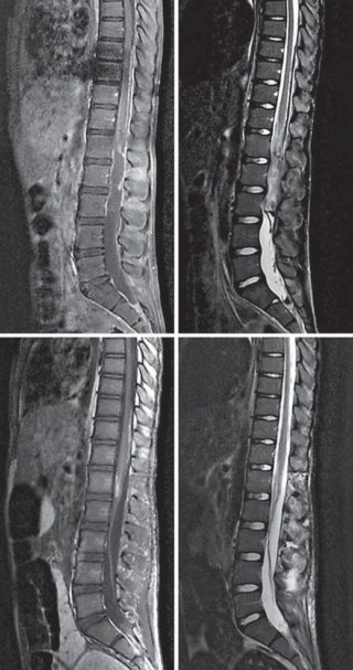
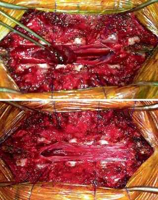
The patient was submitted to thoracic column and lumbar magnetic resonance imaging (MRI) ( fig. 1 ), which gave evidence of an expansive process (heterogeneous and intradural) located between L1 and L3. He underwent emergency surgery: microsurgical excision of the medullary expansive process via laminectomy at L1–L3 and laminoplasty. Once the dura mater had been opened, an extensive hematoma attached to the expansive process was noted ( fig. 2 a).

The tumor lesion presented a cleavage plane of the surrounding structures; however, it was closely attached to the filum terminale, from which it was completely dissected ( fig. 2 b). In a control MRI performed 24 h after surgery, no residual lesion was detected ( fig. 1 c, d). Tomography of the lumbar column performed postoperatively
demonstrated preservation of the thoracic-lumbar curvature and patency of the laminoplasty. Histopathological and immunohistochemical results discovered a myxopapillary ependymoma of WHO grade I.

The patient has improved significantly as to motor deficits; clinical examination 2 months postoperatively revealed proximal and distal muscular strength of grade IV in the RLE, and grade III in the proximal LLE and grade I in the distal LLE. Sensitivity was preserved in the RLE but reduced in the LLE. Reflexes were still absent in both legs. He is able to walk with a walking aid.
Dr. Rodrigo Becco
Médico neurocirurgião graduado pela Universidade de São Paulo (USP), em 2007. Residente em Neurocirurgia pela Santa Casa de São Paulo, em 2014. Mestre pelo Instituto de Assistência Médica do Servidor Público Estadual, em 2015. Membro da Sociedade Brasileira de Neurocirurgia desde 2014.

